Papers summary
- ClabbersGrueneAbrahams2017 : **Clabbers, M. T. B., Genderen, E. Van, Wan, W., Wiegers, E. L., & Gruene, T. (2017). Protein structure determination by electron diffraction using a single three-dimensional nanocrystal. Acta Crystallographica Section D, 73, 738–748. doi
- LatychevskaiaAbrahams2019 : Latychevskaia, T., & Abrahams, J. P. (2019). Inelastic scattering and solvent scattering reduce dynamical diffraction in biological crystals. Acta Crystallographica Section B: Structural Science, Crystal Engineering and Materials, 75, 523–531. doi
- SubrimanianSpence2015 : Latychevskaia, T., & Abrahams, J. P. (2019). Inelastic scattering and solvent scattering reduce dynamical diffraction in biological crystals. Acta Crystallographica Section B: Structural Science, Crystal Engineering and Materials, 75, 523–531. doi
- CowleyMoodie1957 : Cowley, J. M., & Moodie, A. F. (1957). The scattering of electrons by atoms and crystals. I. A new theoretical approach. Acta Crystallographica, 10(10), 609–619. doi This is the original paper on the multislice method.
-
oleynikov2007 : Oleynikov, P., Hovmöller, S., & Zou, X. D. (2007). Precession electron diffraction: Observed and calculated intensities. Ultramicroscopy, 107(6–7), 523–533. doi
ClabbersGrueneAbrahams2017
Protein structure determination by ED of 3D nanocrystal(Experimental) : dimeric polymorph hen egg-white lyzozyme (Diffracting volume ) using Molecular Replacement from monomeric polylalanine model and standard X-ray protein crystallography software.
The claim of a successful solution despite subramanianspence2015 is due to lack of inelastic modelling in multislice simulation. This is backed up by the fact that Friedel pair symmetry is not more violated than for standard X-ray data.
| Software/Method | description |
|---|---|
| Data acquisition | |
| TEM | Talos Arctica/Titan Krios, Parallel beam rotation |
| detector | Timepix pixel( each) |
| Data processing | XDS |
| Structure solution | |
| XSCALE | scaling |
| POINTLESS | MTZ format conversion |
| AIMLESS | merging |
| TRUNCATE | Structure factor amplitudes |
| CHAINSAW | model creation |
| PHASER | Molecular replacement |
| Buccaneer/REFMAC5 | Side chains placement |
| COOT | fitting missing residues |
| Refinement | |
| REFMAC | EXPDTA ELECTRON CRYSTALLOGRAPHY, SOURCE ELECTRON MB flags |
| SFTOOLS | non measured observation removal |
| FREERFLAG | unique test sets creation |
| Micrograph (0.2x0.5x1.4nm) | Diffraction data | Fo vs Fc |
|---|---|---|
 |
 |
 |
LatychevskaiaAbrahams2019
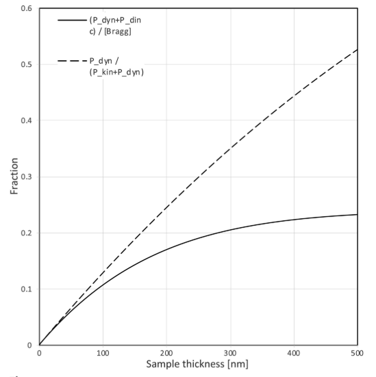
Inelastic scattering and solvent scattering significantly mitigate the effect of dynamical diffraction.
| Probabilities of scattering events | Inelastic fraction of dynamical scattering | Friedel symmetry violation solvent scattering |
|---|---|---|
 |
 |
 |
SubramanianSpence2015
Strong dynamical diffraction prevents single scattering approximation based techniques at crystal thickness above 100-200nm.
Multislice details (ZMULT package) :
- lysozyme Tetragonal space group ( here or here), a=b=79.1A, c=37.8A.
- Structure factor from experimentally refined RHF Doyle-Turner data (H not included)
- 90 beams at 200keV
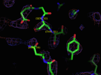
| Friedel symmetry violation | Omit density map : threshold error 37% | MR distinguishable error limit 34% | Thickness limit |
|---|---|---|---|
 |
 |
 |
 |
Oleynikov2007
Comparison of multislice and exp for precessionED and SAED.
| figures | description |
|---|---|
 |
Ewald radii during precED. are intersections with ZOLZ and HOLZ. |
 |
Integration path of the excitation error during precession ED where with azimuthal angle. Oscill is the kinematic rocking curve function where |
 |
integration correction factor where for h=1..30 |
 |
Reflections for azim=0 with forbidden reflections h00 |
 |
Reflections for azim=90 with forbidden reflections 0k0 |
 |
Rfactor for precession ED and SAED run with multislice compared with experimental data in the [001] direction for . precED simus were run for 28 patterns with thickness in range(60,600,20) each of them made of 250 diffraction patterns. |